Structure and Function of the Spinal Cord
The spinal cord is a crucial part of the central nervous system (CNS) that runs from the brainstem down the vertebral column (spine). It plays a vital role in transmitting information between the brain and the rest of the body. Here’s a breakdown of its structure and function:
Structure of the Spinal Cord
-
Length and Location:
- The spinal cord extends from the medulla oblongata at the brainstem to the lumbar vertebrae (around the level of the second lumbar vertebra in adults).
- It is housed within the vertebral column and protected by meninges (layers of connective tissue) and cerebrospinal fluid (CSF).
-
Segments:
- The spinal cord is divided into 31 segments:
- 8 cervical
- 12 thoracic
- 5 lumbar
- 5 sacral
- 1 coccygeal
- Each segment corresponds to a pair of spinal nerves that emerge from the cord and innervate specific regions of the body.
- The spinal cord is divided into 31 segments:
-
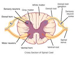
Gray Matter:
- Located in the center of the spinal cord in the shape of an H or butterfly when viewed in cross-section.
- It contains neurons’ cell bodies and is responsible for processing information.
- Anterior (ventral) horns: motor neurons that control voluntary muscles.
- Posterior (dorsal) horns: sensory neurons that receive information from the body.
- Lateral horns (found in thoracic and lumbar regions): autonomic motor neurons involved in the sympathetic nervous system.
-
White Matter:
- Surrounds the gray matter and is made up of myelinated axons that carry signals to and from the brain.
- It is organized into ascending tracts (sensory) and descending tracts (motor).
-
Central Canal:
- A small canal in the center of the spinal cord filled with cerebrospinal fluid (CSF).
-
Spinal Nerves:
- Each segment of the spinal cord gives rise to a pair of spinal nerves. These nerves exit the vertebral column through openings called intervertebral foramina.
- Sensory neurons enter the spinal cord through the dorsal root, while motor neurons exit via the ventral root.
Function of the Spinal Cord
-
Transmission of Signals:
- The spinal cord serves as a conduit for sensory and motor signals between the brain and body.
- Sensory information (e.g., touch, temperature, pain) from the body is carried up the spinal cord to the brain.
- Motor commands from the brain are transmitted down the spinal cord to muscles and glands.
-
Reflex Actions:
- The spinal cord plays a key role in the reflex arc, a quick, involuntary response to stimuli that doesn’t require the brain’s involvement.
- Reflexes help protect the body (e.g., pulling away from a hot object).
-
Coordination of Movement:
- The spinal cord coordinates voluntary movements by transmitting commands from the brain to muscles.
- It also helps in posture control by maintaining muscle tone and balance.
-
Autonomic Functions:
- The spinal cord is involved in autonomic functions, such as regulating heart rate, blood pressure, and digestion, through the sympathetic and parasympathetic nervous systems.